SEM/EDX

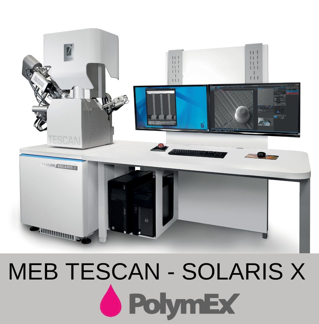
Equipment : Scanning Electron Microscope - SOLARIS X and SEM-FIB Plasma/Gallium +EDX-EBSD
Detection modes available :
- SE: Secondary electrons with topography contrast.
- GSE: Secondary Electron Gas (presence of water vapour).
- BSE: Backscattered electrons with chemical contrast (heavy elements in light on the BSE images)
Sample : 8" max., 130 mm X & Y
Resolution : 0.5nm@30kV
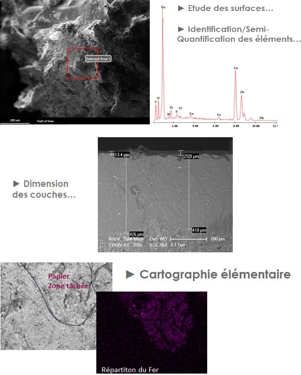

Application domain

- Surface study (defects ...),

- Polished section study (load sizing, layer thickness, etc.),

- Characterization of materials (elemental analysis of all elements) ...

- Biology: Until now, the vacuum constraints imposed by scanning electron microscopy (the electrons must be accelerated under a high vacuum) constituted a handicap for many applications. For example, biological samples, hydrated, oily or insulating products that require observation in their natural state.

Identification et quantification par MEB/EDX

Principle

A SEM (Scanning Electron Microscope) uses an electron beam to obtain enlarged images of very good resolution. It can also be used to identify elements present in the sample thanks to their RX emission property in contact with electrons.
This is called energy dispersive microanalysis (EDS), in which the X emission is processed electronically. The energies of the peaks present are determined and they are automatically compared with an X emission file of known energies.
The analysis is qualitative but we can have access to relatively precise elementary contents.
The analyzes are carried out on raw samples or after metallization (increase in sensitivity).
The element detection threshold varies according to the analysis mode, it is around 500 ppm (0.05% w).

Benefits

- Allows a (qualitative) detection of elements from Carbon to Uranium (> 0.05% w)
- Non-destructive technique,
- Requires little sample (a few µg),
- No sample preparation

Examples

OUR ANALYTICAL EQUIPMENT 

During injection into the GC, all organic compounds will be separated by the chromatographic column and then identified by the MS (mass..
GCMS-FID

GCMS-FID

The sample is directly injected into the pyrolyzer oven heated to 550 ° C using a solid syringe. This instantly causes the fragmentation of..
PYROLYZER

PYROLYZER

During the injection into the LC, all the semi-volatile organic molecules will be separated by the chromatographic column and then detected..
UPLC/UV/TOF-MS

UPLC/UV/TOF-MS

During the injection into the LC module, all the semi-volatile organic molecules will be separated by the chromatographic column and then..
UPLC-MS-MS

UPLC-MS-MS

The HeadSpace / GCMS technique does not require preparation. The sample is placed in a gas tight vial and hermetically sealed using a..
HEADSPACE/GCMS

HEADSPACE/GCMS

This spectral analysis makes it possible to know the nature of the covalent bonds present in the sample. Each of them (for example..
IRTF SPECTROMETER

IRTF SPECTROMETER

A SEM (Scanning Electron Microscope) uses an electron beam to obtain enlarged images of very good resolution. It can also be used to..
SEM/EDX

SEM/EDX

CENTRIFUGE

CENTRIFUGE

Certain molecules form by reaction colorimetric complexes having maximum absorption in UV-Visible. The color of these complexes is read..
UV-VIS SPECTROMETER

UV-VIS SPECTROMETER